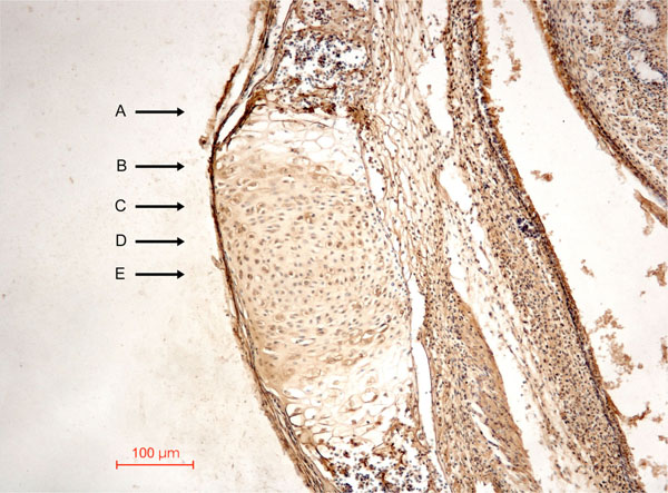
Fig. (1) Photomicrograph of a synchondrosis in neonatal mice showing different zones across one side of the synchondrosis. (A) Zone of
vascular erosion and invasion. (B) Zone of chondrolacunar hypertrophy and matrix calcification. (C) Zone of matrix production or
matrixogenic zone. (D) Zone of cellular proliferation. (E) Central zone or resting zone.