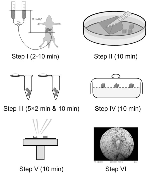
Fig. (1) Scheme depicting the different steps involved in the preparation of liver tissue for ultrastructural scanning electron microscopy investigation. Step I, perfusion-fixation of the liver with 1.5% glutaraldehyde; Step II, tissue slicing in blocks of 1 mm3; Step III, submerging tissue blocks in an organic reagent with a fast evaporation rate; Step IV, air-drying in a desiccator; Step V, mounting of the samples on stubs and sputter-coating; and Step VI, imaging within a table-top scanning electron microscope