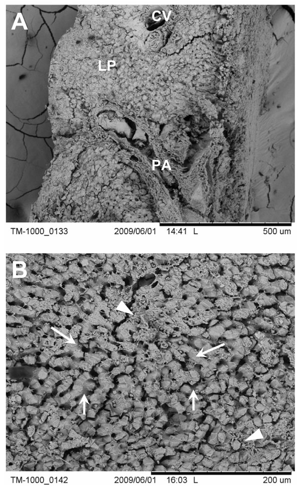
Fig. (2) Low (A) and intermediate (B) magnification of liver tissue. (A) PA, portal area; CV, central vein; LP, liver parenchyma. (B) Large arrows denote the hepatocytes that make-up the liver parenchyma and the small arrows indicate the microvascular bed or sinusoids. Arrowheads, extracellular matrix components.