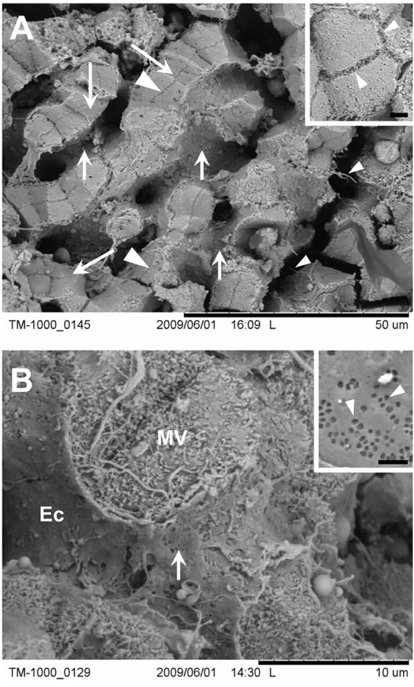
Fig. (3) The ultrastructure of liver tissue as seen at high magnification. (A) The sinusoids are readily visible (small arrows) and are surrounded by hepatocytes (large arrow). Large arrowhead, bile canaliculi; small arrowhead, extracellular matrix components. Inset depicts a detailed view of bile canaliculi (arrowheads). Scale bar, 1µm. (B) Investigation of the sinusoidal lumenwhich is delineated by the endothelial lining (Ec)shows the presence of fenestrae (arrow). Note also the numerous microvilli projecting from the hepatocytes (MV). Inset shows fenestrations (arrowheads) recorded at the highest possible magnificationi.e. 10,000×with a desktop scanning electron microscope. Scale bar, 1µm.