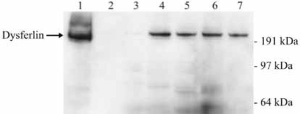
Fig. (5) Confirmation of the immunoprecipitation of dysferlininteracting
partners. Lane 1 shows the level of dysferlin in the
lysate before immunoprecipitation. As controls no lysate were
added to the beads (lane 2) or the lysate was not pre-incubated with
any antibody (lane 3). For the immunoprecipitation antibodies
against the following proteins were used: dysferlin (lane 4), adaptin
alpha (lane 5), striatin (lane 6) and utrophin (lane 7).