- Home
- About Journals
-
Information for Authors/ReviewersEditorial Policies
Publication Fee
Publication Cycle - Process Flowchart
Online Manuscript Submission and Tracking System
Publishing Ethics and Rectitude
Authorship
Author Benefits
Reviewer Guidelines
Guest Editor Guidelines
Peer Review Workflow
Quick Track Option
Copyediting Services
Bentham Open Membership
Bentham Open Advisory Board
Archiving Policies
Fabricating and Stating False Information
Post Publication Discussions and Corrections
Editorial Management
Advertise With Us
Funding Agencies
Rate List
Kudos
General FAQs
Special Fee Waivers and Discounts
- Contact
- Help
- About Us
- Search


The Open Paleontology Journal
(Discontinued)
ISSN: 1874-4257 ― Volume 5, 2014
Nano-Scale Spheroids and Fossils from the Ediacaran Doushantuo Formation in China
Tenger Borjigin1, Leiming Yin2, *, Lizeng Bian3, Xunlai Yuan2, Chuanming Zhou2, Fanwei Meng2, Xiaomin Xie1, Fang Bao1
Abstract
Exceptionally preserved nano-scale spheroids derived from microbial processes and nano-scale fossils have been discovered from the black shales of the Jijiawan section of the Ediacaran Doushantuo Formation in the Yangtze Gorge area of Hubei Province, southern China. The numerous soccer ball-like spheroids are pyritized. Their morphology and abundant preservation may suggest that they could possibly be related to larger spheroids, regardless of the tremendous dimensional gap found in the phosphorite and cherts of the Doushantuo Formation, including those recognized as ‘embryos’. The colony-like spheroids preserved in situ and obtained by acid maceration are compared with known Neoproterozoic microfossils—Bavlinella faveolata (or Sphaerocongregus variabilis). Additionally, nano-scale fossil bodies, characterized by morphological features comparable to living cyanobacteria, fungi and possible unicellular heterotrophic protists were observed in different minor laminae of the black shale samples. This study aims to reveal the aspects of nano-scale biota preserved in the black shale of the Ediacaran Doushantuo Formation, and highlight the taphonomy of microorganisms during the key transition from the anoxic deeper oceans to the oxygenated oceans of the early Ediacaran interval.
Article Information
Identifiers and Pagination:
Year: 2014Volume: 5
First Page: 1
Last Page: 9
Publisher Id: TOPALOJ-5-1
DOI: 10.2174/1874425701405010001
Article History:
Received Date: 19/11/2013Revision Received Date: 20/12/2013
Acceptance Date: 2/1/2014
Electronic publication date: 24/1/2014
Collection year: 2014
open-access license: This is an open access article licensed under the terms of the Creative Commons Attribution Non-Commercial License (http://creativecommons.org/licenses/by-nc/3.0/), which permits unrestricted, non-commercial use, distribution and reproduction in any medium, provided the work is properly cited.
* Address correspondence to this author at the State Key Laboratory of Palaeobiology and Stratigraphy, Nanjing Institute of Geology and Palaeontology, Chinese Academy of Sciences, Nanjing 210008, Jiangsu, China; Tel: 0086-025-83282240; E-mail: lmyin2012@gmail.com
| Open Peer Review Details | |||
|---|---|---|---|
| Manuscript submitted on 19-11-2013 |
Original Manuscript | Nano-Scale Spheroids and Fossils from the Ediacaran Doushantuo Formation in China | |
1. INTRODUCTION
Based on geochemical and molecular palaeontological research, the Neoproterozoic Ediacaran period has been considered a key transition point in the geological history of the Earth [1-3]. Ediacaran fossil discoveries, including the first appearance of large and diverse soft-bodied organisms, basal unicellular protists (acanthomorphic acritarchs), animal embryos and various seaweeds, indicate that the members of the Ediacaran biota arose shortly after the last Neoproterozoic glaciation. This evidence suggests that there was a causal link between their evolution and the oxygenation event [1, 2, 4]. Geochemical data indicate that oxygenation of the deep ocean happened in the mid-Ediacaran period [3-7]. Thus, it seems likely that atmospheric oxygen was a key factor in the development of Ediacaran fauna. However, as yet, no compelling fossil record has been produced as evidence of the biota for the early Ediacaran relatively anoxic, ferruginous ocean.
1. GEOLOGICAL SETTING
The 635–551 Ma Ediacaran Doushantuo Formation [8] is sandwiched between the underlying Nantuo tillites and the overlying dolomites of the Dengying Formation in southern China. Since the 1970s, many significant permineralized fossils, including acanthomorphic acritarchs, eukaryotic algae, animal embryos, diapause egg cysts, lichen and possible microscopic animals have been found in the cherts and phosphates of the Doushantuo Formation [9-15].
Here, we report on a new discovery of nano-scale fossils, and nano-scale spheroids derived from microbial processes, preserved within the black shales of the Jijiawan section of the Ediacaran Doushantuo Formation in the Yangtze Gorge area of Hubei Province, South China.
The Jijiawan section is located on the western limb of the Huangling Anticline in the Yangtze Gorges area (30°52′54.4″N, 110°52′ 38.2″E) (Fig. 1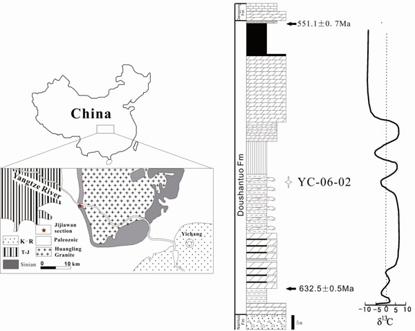 ). According to geochemical data acquired for the Jijiawan and other sections in the Yangtze Gorge area [6, 16, 17], the black shale bed contains abundant organic matter [ω(Corg)% = 3.98] and is characterized as a horizon that contains a notable negative peak value of carbonate (δ13Ccarb) (Fig. 1). The shale samples are positioned just beneath beds containing abundant and diverse acanthomorphic acritarchs, red algae and micrometazoans found from other sections of the Doushantuo Formation [6, 18, 19]. The black shale bed has a likely age of about 600 Ma, based on a 632.50±0.48 Ma U-Pb zircon dating for the ash bed lying 9.5 m above the base of the Doushantuo Formation, a 550.55±0.55 Ma U-Pb zircon dating for the ash bed in the uppermost Doushantuo Formation in the studied section [8], and adopting the reference of a 614.0±7.6 Ma zircon SHRIMP U-Pb age for the mid-Doushantuo Formation in the Zhangcunping section of the Yangtze Gorge area [20].

3. METHODS
The black shale samples studied here were collected from a horizon 70 m above the base of the Doushantuo Formation in the Jijiawan section.
Nano-scale spheroids and fossils were preserved in situ and examined using SEM (performed on a LEO 1530, JSM 6300 at the Nanjing Institute of Geology and Palaeontology and Wuxi Research Institute of Petroleum Geology) on fresh samples of the black shale that were either left uncoated or coated with gold (gilded). They occur in different minor laminae, which are characterized either by abundant pyrite or the presence of distinct gypsum minerals. The uncoated and gilded specimens were used to determine elemental composition using a computerized energy-dispersive X-ray micro-analyser system (EDX). In addition, some specimens preserved in situ were analysed by Raman spectroscopy (performed on a Renishaw in via at the Wuxi Research Institute of Petroleum Geology) to provide information on their material components and molecular structure. The measurements were performed on polished rock specimens to determine Raman spectra for both nano-scale spheroids and adjoining pyrite framboids.
To further characterize the observed spheroidal structures and nanofossils under SEM, rock samples were macerated with diluted hydrochloric and hydrofluoric acids to obtain organic residues. Some uncoated specimens, which were selected from organic residues, were analyzed for their elemental composition using EDX.
4. RESULTS
In the study, nano-scale spheroids obtained by acid maceration from the sampled black shale of the Doushantuo Formation and observed under SEM could be represented by individual and colony-like forms. Individual forms include soccer ball-like spheroids characterized by polygonal cracks, and simple spheroids showing sub-spherical to spherical aspects that normally display wall-like vesicles and hollow interiors. These are all preserved in situ with an amorphous aggregation or scattered distribution (Fig. 2A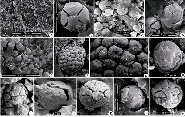 ).
).

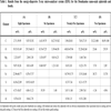
The soccer ball-like spheroids are embedded in the membraniform clay minerals of the host rock and preserved in situ together with a few pyrite framboids. Most spheroids were nearly the same size (4–5 μm in diameter, n = 250), but some were smaller (3 μm, n = 60) and the smallest diameter was < 1 μm (n = 25). Single spheroid specimens commonly had about eight scalene pentagon-hexagon (Fig. 2B–E, L) or irregular polygonal partitions (Fig. 2M) cleaved by grooves. A few specimens display tuberculiform ornamentation on their surfaces (Fig. 2I). Even though all of the spheroids contain distinctively high amounts of carbon and organic matter (based on EDX spectra (Fig. 4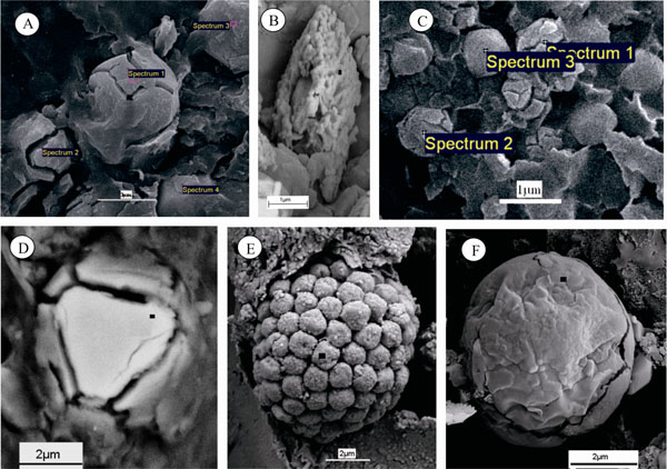 , Table 1) and Raman spectra (Fig. 5
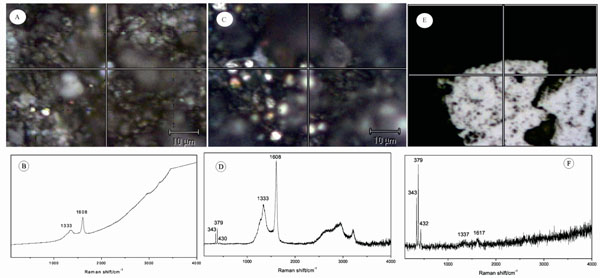 ), they showed pyritization to different degrees, or sometimes merely ferrugination and sulfuration. Their Fe/S ratios varied from 3:1 to nearly 45:1 (see Table 1C), while containing both iron and sulfur.


A few solitary and slightly larger spheroidal specimens (Fig. 2K, J), preserved together with the soccer ball-like spheroids in the same minor laminae or laminae-bearing gypsum minerals, displayed similar pyritization and sulfuration. A few specimens (Fig. 2H) showed weak sulfuration or other mineralization containing calcium or silicon in their EDX spectra.
Another notable colony-like form has been obtained from organic residues of acid maceration and observed under SEM. These specimens (Fig. 2F; Fig. 4E) resemble pyrite framboids in their rough morphology, but they display cell-like units rather than pyrite crystals or pyrite spheres, because of their plastic ‘cell walls’ and empty interiors (Fig. 2F, G). The EDX spectra of these specimens shows that the element contents are different, which indicates that they have weak ferrugination or sulfuration and no remarkable mineralization, only containing carbon, oxygen and other minerals (Fig. 4I). Moreover, the fact that such specimens (Fig. 2F) were obtained by acid maceration, might be implied by the nature of their acid resistant organic walls.
In addition, some nano-scale morphological bodies have been discovered under SEM in minor lamina containing gypsum minerals within the black shale sample. Based on their physical features, jar-like and oblate morphological forms can be recognized. The jar-like specimens (Fig. 3D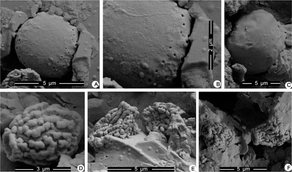 , E, F), 4–7 µm in length and 3–5 µm wide (n = 12), display a horn-like circular loop at one end of the ellipsoidal body, exhibiting lengthways ridged thickenings (or ‘ornamentation’). We assume that they are hollow, because some observed specimens were compressed (Fig. 3E). They normally appear individually, with only occasional connections between one another. Their EDX spectra show either weak ferrugination containing a small amount of sulfur or show only the curved peak of carbon.

The oblate body, ringed with tens of minute holes, was about 5 µm in size and was embedded in gypsum crystals and clay minerals (Fig. 3A, C). Under high-powered SEM, the minute holes display different dimensions (Fig. 3B) distributed at distances of 300–500 nm. Some of the slightly larger holes had rising circular aperture edges. Only one such specimen is confirmed; other similar specimens are questionable because they do not show the minute holes (Fig. 3C).
5. DISCUSSION
Based on studies of lithology, biostratigraphy and geochemistry, the Ediacaran Doushantuo succession in the Yangtze Gorges area is inferred to be deposited on a rimmed carbonate shelf with a shelf-margin barrier separating the shelf lagoon from the open ocean [21]. The Doushantuo Formation was deposited at greater depths in the intra-shelf basin during the same geological period [18]. The studied samples were collected from interbedded organic-rich black shale of bedded dolostone from the lower part of the Jijiawan section, where there is an obvious, but small, negative peak in δ13C. Compared with adjacent sections, such as the Miaohe and Jiulongwan sections in the Yangtze Gorge area, the depositional environment of the sample should lie near the transition from anoxic/euxinic deeper water to oxygenated shallower water [21, 22]. The presence of variable minor laminae containing either pyrite or gypsum might correspond to fluctuations in the environment of deposition during the geological time interval. This supports the interpretation of a stratified redox model for the Ediacaran Ocean where it is observed in the Nanhua Basin [22, 23]. The fact that the minor laminae containing abundant nano-scale pyritized spheroids and framboidal pyrite changed immediately (< 0.5 mm thick) in the above minor laminae, with gypsum, preserving nano-scale unicellular fossils, indicates that the depositional environment likely was euxinic near the water-sediment interface. This interpretation is supported by both the known fossil record of the Ediacaran Lantian Formation in Anhui, China [24], and the study of framboidal pyrite in modern sediments [25]. The studied nano-scale spheroids and fossils were preserved in black shale formed in redox conditions.
Nano-scale spheroids of the Doushantuo Formation, especially those presenting as soccer ball-like spheroids, are distinguished from normal pyrite crystals, pyrite spheres similar to those reported from Devonian black shales in North America [26] or euhedral crystals in external morphology [27]. They only occur in minor laminae with pyrite framboids or pyrite crystals and indicate a transition from sulfuration, ferrugination to pyritization, based on the analysis of EDX spectra (Table 1). Such specimens might represent microbiotic remains that were altered mainly by pyritization. This interpretation can be supported by Raman spectra analysis (Fig. 5C). Although the measured soccer ball-like spheroid shows characteristic Raman spectra curves of pyrite (FeS2) at the 343, 379 and 432 cm-1 bands, it displays remarkable Raman point spectra at the 1333 and 1608 cm-1 bands and three wider and gently curved peaks between 2500 and 3250 cm-1 bands (Fig. 5C, D), which are recognized as having characteristic Raman spectral peaks for carbonaceous (organic) matter [28]. The other isolated spheroid specimen (Fig. 5A) only produced 1333 and 1608 cm-1 bands for carbonaceous (organic) matter (Fig. 5B). In contrast with typical Raman spectra of pyrite framboids or crystals (Fig. 5E, F) preserved in the same minor laminae within a rock sample, spheroids with polygonal cracks are more strongly recognized to have originally been microorganisms, rather than abiotic structures.
In morphological style, regardless of the tremendous dimensional gap, the ‘soccer’ globules (Fig. 2B, C, D, L, M) are quite similar to some Doushantuo phosphatic mophological forms – particularly Parapandorina, Spiralicellula and Caveasphaera – which have been recognized as animal cleavage embryos [11, 29]. However, present nano-scale specimens do not show distinct ‘vesicle walls’ and represent a fixed form as a series of cracks. The differences might be the result of varied fossil preservation. So far, well-preserved animal embryo fossils of the Ediacaran Doushantuo Formation are preserved in either phosphatization or silicification in a few later strata. In modern marine embryos, oxygen and nutrition in aquatic environments are important limiting factors to influence development of embryos [30, 31]. Though the studied nano-scale spheroid specimens are so tiny size for imagining as embryos, the anoxic/euxinic deeper water could be restricted their development. However, the studied nano-scale spheroid specimens were deformed by pyritization during burial preservation. Additionally, the tiny size makes the evaluation of their interior structures very difficult for detailed study on a subcellular level.
In modern microorganisms, a giant sulfur bacterium — Thiomargarita — discovered in Namibian Shelf sediments shows similar globules during cell reductive division [32, 33]. However, there are distinct differences between them. The present globules are much smaller than the giant Thiomargarita and no series of reductive cell division like those of Thiomargirita appears in the present spheroids. Furthermore, precipitation of phosphorous minerals, which is normally mediated by Thiomargarita [32], has not been found in associating spheroids and even in the studied black shale sample.
In external morphology, the ‘soccer’ globules look like the spore of living Licea eremophila [34] of the myxomycetes. However, the latter displays raised bands on the surface rather than cracks, and moreover, no sporocarp-like remains have been found from the studied rock samples.
Although the cause of polygonal cracks in the spheroid specimens remains uncertain, their morphology and ready preservation may suggest that nano-scale spheroids preserved in black shale could relate to larger spheroids found in the upper part of the Doushantuo Formation, including those recognized as ‘embryos’.
Specimens characterized by isolated spheroids (Fig. 2H, J, K) can be compared with some known coccoid microfossils reported from Proterozoic cherts and black shales in China and elsewhere in the world. For example, one form—Nanococcus vulgaris—described by Oehler [35] from the Y.C. Pyritic Shale Member of the Barney Creek Formation, McArthur Group, Australia, is similar to studied isolated spheroids in that they may be characterized by small (normally < 10 µm) isolated spheroids and their loose distribution. Nanococcus vulgaris has been compared with planktonic members of the modern chroococcacean genus Aphanocapsa Nageli [35], but studied isolated spheroids seem to lack an amorphous organic matrix as colony preservation of Aphanocapsa .
The specimens of colony-like spheroids (Fig. 2F) appear to be distinguished from abiotic pyrite framboids [24, 36, 37] and possible greigite framboids [26] by the fact that they display cell-like units, exhibiting relatively larger sizes and plastic characters similar to organic matter deformed or ruptured by compression (Fig. 2F, G). Previously, distinguishing biotic from abiotic processes in the formation of pyrite framboids has not been well understood. The identification of the potential signatures of such activities requires improved understanding of pyrite chemical- and bio-oxidation mechanisms [38]. Among studied specimens, rare ferrum or sulfur and high carbon elements were detected with EDX for some specimens. However, a few specimens obtained by acid maceration (Fig. 2F) only show elemental carbon and oxygen in their EDX spectra. Therefore, such colony-like spheroids may originally have been endosporous microorganisms that were widely distributed in deeper weakly reducing and oxidic conditions, such that they ended up in different taphonomic circumstances.
Similar morphological microfossils, such as Bavlinellafaveolata or Sphaerocongregusvariabilis, which was interpreted as resembling endosporagia of cynobacteria [39], often occur within extraordinarily low-diversity acritarch assemblages obtained from late Neoproterozoic sediments from ecologically stressed palaeoenvironment [39-42]. The studied specimens are consistent with Bavlinella faveolata with regard to morphological features. As Knoll and Blick (1981) [41] concluded, Bavlinella faveolata represents biological organization different from pyrite framboids on the basis of their organic composition and discrete time range.
The nano-scale jar-like bodies ornamented with ridge-like protuberances (Fig. 3D, E, F) seem to have been discovered first in the Ediacaran Doushantuo Formation of South China.
Previously, vase-shaped forms of microfossils had been discovered in Neoproterozoic sediments in China and elsewhere in the world. Those fossils are characterized by vase- or tear-like shaped tests, consisting of a rounded pole gradually tapering toward a “neck” that ends in a single aperture; their systematic affinity has been suggested to be similar to that of testate amoebae [43]. Present jar-like bodies, although also displaying a similar jar- or ‘vase’-shape, differ from vase-shaped microfossils in having a horn-like annulation at one pole and ridge-like protuberances on their surface. Thus, the jar-like bodies, at present, cannot be referred to simply as vase-shaped microfossils with an affinity to amoebae. Additionally, based on hollow cavities found in the jar-like bodies, the morphology of the bodies is similar to fungal spores [44].
In the minor gypsum mineral-bearing laminae, the occurrence of specimens containing an oblate body with two or three rows of numerous minute holes or with no obvious ornamentation (Fig. 3A, C) could imply that the alteration of nano-scale fossil association was accompanied by changes in depositional and taphonomic circumstances from deeper reducing conditions to shallower evaporative conditions. So far, only one specimen bears numerous minute holes. These holes are presumed to have been preserved as primitive structures, because they are unlikely to have formed by secondary diagenetic or taphonomic distortion and such structures could not be the result of mineral dissolution, since the studied specimen is preserved in situ in black shale rather than extracted by acid maceration. Based on their rather simple smooth oblate-spheroid morphology, the specimens are comparable to unicellular algae. However, the observed morphological character with numerous minute holes along its margin is not known in either fossil records or living forms of unicellular algae. Therefore, the presence of the minute holes could be traces of a biological function such as breathing holes or some receptacle for moving structures like cilia. The former case may be a more reasonable interpretation because in that case it could remain as a unicellular heterotrophic protist. However, the hypothesis requires confirmation using similar specimens and supplemental evidence.
Although the above comparisons and discussion of the nano-scale fossils or spheroids are based mainly on morphology and the analysis of limited elemental compositions and organic material, they may enhance our views on the diversity of life and the mechanisms of its preservation at the time of the early Ediacaran anoxic, ferruginous ocean.
6. CONCLUSIONS
Morphological features similar to those of modern cyanobacteria characterize the preservation of nano-scale fossils [45]. Possible fungal spore and unicellular heterotrophic protists may imply that a diverse nano-scale biota, including prokaryotes and heterotrophic eukaryotes, existed in the early Ediacaran anoxic deeper ocean. Although most of the nano-scale spheroids and fossils obtained from the black shale of the lower part of the Doushantuo Formation were strongly carbonized and mineralized, these findings further reveal aspects of life during this key period of transition from anoxic deeper oceans to oxygenated oceans.
Consequently, the investigation of the biological link that may have existed between such nano-scale microbes and much larger microfossils, particularly acanthmorphic acritarchs, animal embryos and other possible animal remains, during the earlier Ediacaran period is significant. Mineralogically, the studied black shales contain abundant pyrite and gypsum, which alternate within thin laminae. This may signify that redox conditions were fluctuating and a semi-enclosed marine lagoonal palaeoenvironment could have been suitable for the sedimentation and diagenesis of the studied Doushantuo black shales. The fact that many specimens of colony-like spheroids are comparable in their nano-scale biotic associations to Bavlinellafaveolata and other cyanobacteria may suggest that an extraordinarily stressed environment existed during this key geological interval.
CONFLICT OF INTEREST
The authors confirm that this article content has no conflicts of interest.
ACKNOWLEDGEMENTS
We are grateful to Andrew H. Knoll for discussions and significant suggestions during preparation of the manuscript and to two anonymous reviewers for helpful comments. Guoliang Tao and Qigui Jiang are thanked for fieldwork assistance; Fengbao Huang is thanked for palynological maceration. The assistance of Yonxiang Mao, Chunzhao Wang and Zhang Qingzhen with the SEM work is appreciated. The work was supported by the National Natural Science Foundation of China (40839910, 41130209) and State Key Laboratory of Palaeobiology and Stratigraphy (Nanjing Institute of Geology and Palaeontology, CAS (No. Y326150507).