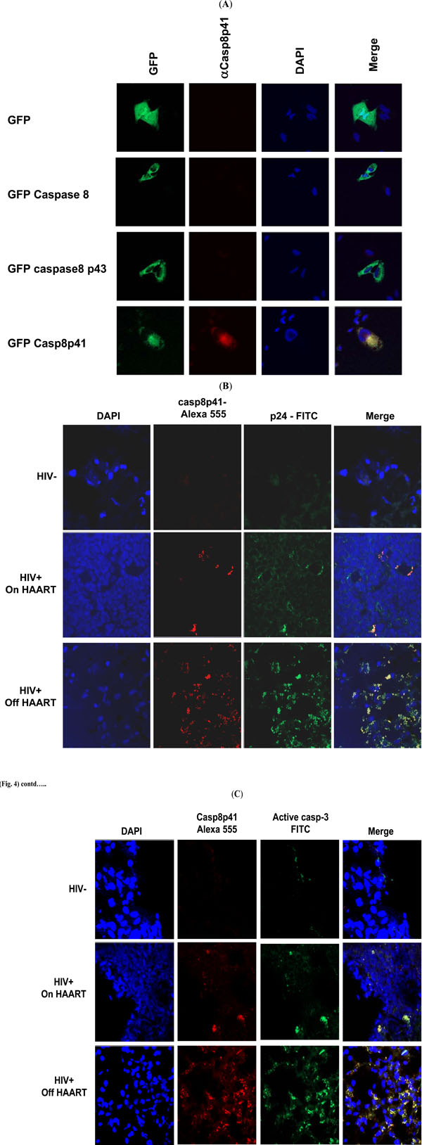
Fig. (4). (A) Hela cells were transfected with the indicated GFP constructs and stained with anti-casp8p41 PE and DAPI. (B) Lymph node sections from HIV negative or HIV infected patients with either suppressed or non-suppressed levels of viral replication were stained with anti-casp8p41 (red) and anti-p24 (green) counterstained with DAPI to identify nuclei and analyzed by confocal microscopy. Results representative of three or more analyses each. (C) Lymph node sections from HIV negative or HIV infected patients with either suppressed or non-suppressed levels of viral replication were stained anti-casp8p41 (red) and anti-active caspase 3 (green) and counterstained with DAPI to identify nuclei and analyzed by confocal microscopy. Results representative of three or more analyses each.