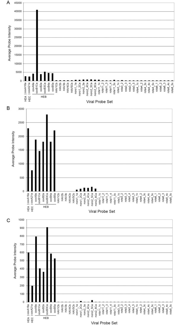
Fig. (7) Comparison of hybridization profiles of three CV strain targets: average probe intensity. Viral genomic RNAs isolated directly from clarified tissue culture supernatants of infected cells were used for RT followed by PCR amplification and labeling prior to array hybridization as described in Materials and Methods. The hybridization data (normalized probe intensities) were converted to average probe intensities [15] and plotted vs each individual probe set following hybridization with either CVB1 (panel A), CVA3 (panel B) or CVA5 (panel C). The underlined identifies the human enterovirus species (HEA, HEB and HEC) represented by a CV probe set.