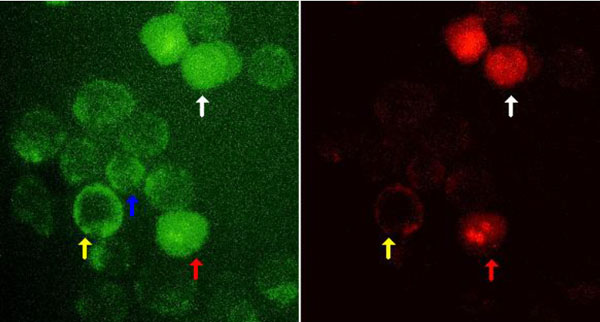
Figure 3 Microphotography of HeLa cells undergoing apoptosis stained by FITC-Dextran and PI (x1000). HeLa cells were incubated at 43,5°C for 1 hour and at 37°C for 8 hours then labeled with FITC-Dextran and PI and analyzed by fluorescence microscopy. The left micrograph shows the green fluorescence (FITC), and the right micrograph shows the red fluorescence (PI). Because FITC-Dextran stained the early and late stages of apoptosis, the cells were labeled with FITC-Dextran more than PI. The white arrow shows apoptotic cells with plasma membrane rupture, the red arrow indicates the apoptotic cells with nucleus fragmentation, the yellow arrow shows apoptotic cells with nucleus alteration, and the blue arrow indicates apoptotic bodies. The living cells impermeable to FITC-Dextran aren't stained.