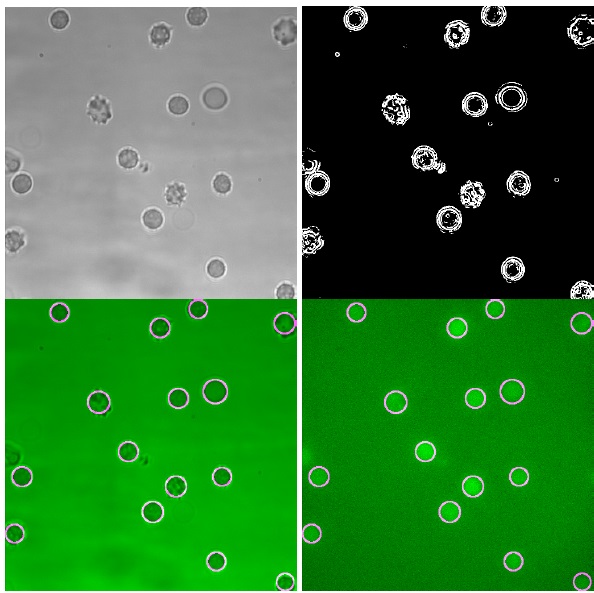
Fig. (3)
Typical sequence of one single processed image from an experiment with the activator A23187. Top left: Original brightfield image. Top right: Fully preprocessed image. Note how sharp edges such as cell boundaries are extracted. Bottom left: Original brightfield image with drawn in circles. Bottom right: Corresponding fluorescence image (stained with Fluo-4) with drawn in circles.