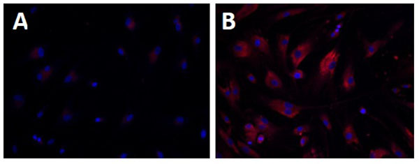
Fig. (1)
Increased nonpolar lipid accumulation in WD fibroblasts detected by Nile-Red dye staining. Nile-Red dye stains for nonpolar lipids including triglycerides and esterified cholesterols that accumulate in WD fibroblasts (B) compared with wild type fibroblasts (A).