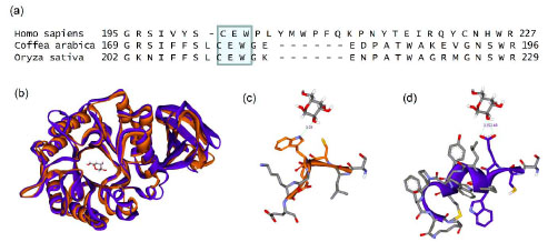
Fig. (7) Comparison of GLA structure and sequence from human, rice, and coffee bean. (a) Extract of a sequence alignment from three species, conserved residues CEW are highlighted. (b) Structural alignment of rice (orange) and human (purple) proteins. A bound galactose molecule is at the center of the figure. Protein folding is conserved between the species. (c) In the rice structure, cysteine 210 and tryptophan 212 (orange) are solvent exposed in close proximity to the bound sugar. (d) However, in the human structure, the same conserved sequence plays a very different structural role with the equivalent cysteine 202 and tryptophan 204 buried and glutamate 203 exposed, proximal to the bound sugar.