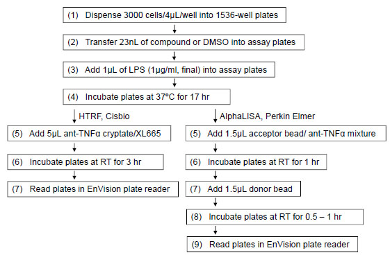
Fig. (2) qHTS protocols for HTRF-based and AlphaLISA-based TNF-α assays. THP-1 cells were dispensed at 3000 cells/well in 1536-well plates. After 23 nL compound or DMSO only was added into the assay plates, 1 µL assay medium with or without 5 µg/mL LPS (final concentration, 1 µg/mL) was added into the assay plates. The assay plates were incubated for 17 hours at 37°C. For HTRF-based assay format, 5 µL anti-TNFα cryptate/XL665 mixtures were added into the assay plates. After the assay plates were incubated at RT for 3 hr, fluorescence intensity (320 nm excitation, 615 and 665 nm emission) was measured using an Envision plate reader (Perkin Elmer). For AlphaLISA-based assay format, 1.5 µL of the mixture containing acceptor beads and anti-TNF-α antibody was added into each well. The assay plates were incubated in the dark at RT for 1 hr, followed by the addition of 1.5 µL of donor bead. After the assay plates were incubated in the dark at RT for another 0.5 to 1 hr, the assay plates were read in the AlphaScreen mode on the Envision plate reader (Perkin Elmer).