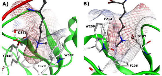
Fig. (3) A) Binding cavity of L3MBTL1 bound to H4K20me2 (PDB:2RJF) and B) Binding cavity of SCML2 bound to a mono-methylated lysine (PDB: 2VYT). Protonated methyl-lysines are displayed in ball and stick model with gray carbon atoms, key binding site residues are displayed in stick model with white carbon atoms.