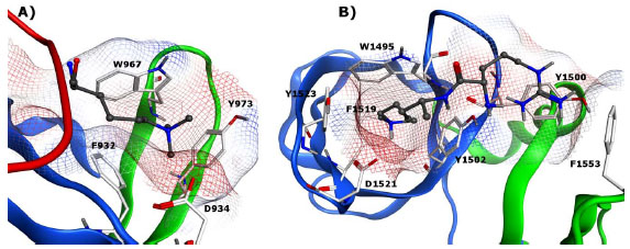
Fig. (6) A) Double tudor domain JMJD2A with the aromatic cavity bound to H3K9me2 (PDB: 2OX0) B) Binding pocket of 53BP1 tandem tudor domain bound to H4K20me2 (PDB: 2IG0). In both structures, the two tudor domains are shown in blue and green, respectively. The trimethyl-lysine in A and the protonated dimethyl-lysine and the neighboring H4R19 residue in B are displayed in ball and stick model with gray carbon atoms. Key binding site residues are displayed in stick model with white carbon atoms.