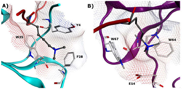
Fig. (7) A) Aromatic binding pocket of CBX5 (human HP1α) bound to H3K9me3 (PDB: 3FDT) B) Chromodomain CHD1 binding pocket with H3K4me3 (PDB: 2B2W). In both structures, the trimethylated lysines are displayed in ball and stick model with gray carbon atoms, key binding site residues are displayed in stick model with white carbon atoms.