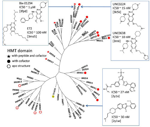
Fig. (1) Structure coverage of human PMTs mapped on a phylogenetic tree. The phylogeny is based on a multiple sequence alignment
of the catalytic domain. Structures of SET domain PMTs are shown in red, arginine PMTs in gray and DOT1L (the only non-SET domain
lysine PMT) in yellow. Ternary complexes with substrate and cofactor are indicated by a star, complexes with the cofactor alone by a full-circle,
and apo-structures by an open-circle. Co-crystallized inhibitors are indicated.