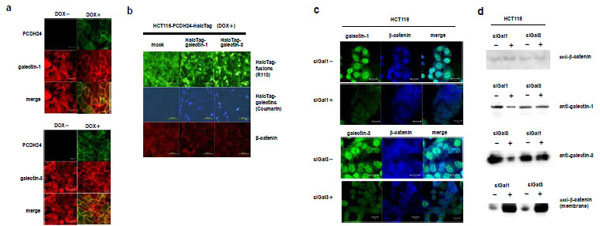
Fig. (3). Effects of galectin-1 and -3 expression on the subcellular localization of β-catenin (a) Subcellular localization of endogenous galectin-1 and -3 in HCT116 PCDH24-HaloTag cells. HCT116 PCDH24-HaloTag cells were cultured with or without DOX for 36 h before formalin fixation. For immunofluorescent analysis, galectins were labeled using specific anti-bodies against galectin-1 or -3 (red). PCDH24-HaloTag was labeled with the HaloTag® TMR ligand (green). The scale bar represents 20 µm. (b) Translocation of β-catenin to the nucleus in PCDH24-HaloTag-expressing HCT116 cells (DOX+) caused by the over-expression of the galectins. Immunofluorescent analysis of β-catenin in the cells in the presence or absence of ectopically-expressed HaloTag-fused galectins was performed using an antibody against β-catenin (blue). PCDH24-HaloTag was fluorescently-labeled using HaloTag® R110Direct™ ligand prior to the transfection of HaloTag-fused galectins expression clones. HaloTag-fused galectins were fluorescently-double stained using HaloTag® R110Direct™ ligand and coumarin ligand. The HaloTag-fused galectin expression clones used here were obtained from the Kazusa Collection of Flexi HaloTag Clones [26]. The cells were fixed at 24 h after the transfection of HaloTag-galectin-1 or HaloTag-galectin-3 expression clones. The scale bar represents 200 µm. (c) Effects of siRNA against the galectins on the subcellular localization of β-catenin. The subcellular localization of the galectins and β-catenin in HCT116 cells was observed in the presence or absence of the ga-lectin siRNAs. Endogenous galectins and β-catenin were labeled with specific antibodies. The scale bar represents 20 µm. (d) Reduction of endogenous galectins and increase in membrane-localized β-catenin by siRNA against galectins. Western blot experiments were performed for whole cell lysate using anti-β-catenin, anti-galectin-1 and -3 and for the membrane fraction using an anti-β-catenin antibody.