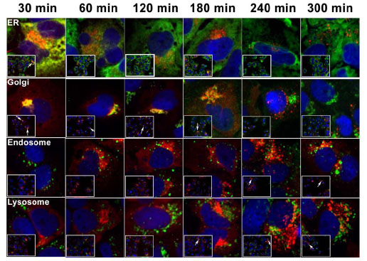
Fig. (3) Subcellular localization of intracellular PCSK9. 14-13-E4 cells were labeled with 5 µM TMR (Red) for 15 min. The unbound ligand was washed off. Cells were fixed (4% Para-formaldehyde, 0.2 M Sucrose in PBS) and permeabilized (0.1% Triton X-100 in PBS) at various time points as indicated. The cells were then counterstained with different subcellular organelle markers (Green), including PDI (ER), Golgin97 (Golgi complex), EEA1 (early endosome), and LAMP1 (lysosome). The images were acquired with an Evotech Opera Confocal High Content Imager with appropriate fluorescence filters. Co-localization of PCSK9 with each organelle is indicated by arrows. Intracellular PCSK9 is primarily located in the ER and Golgi at the time of labeling, and eventually moves to lysosomes. Co-localization of intracellular PCSK9 with early endosomes is observed as well.