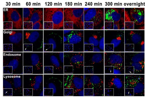
Fig. (4) Subcellular localization of internalized PCSK9. 14-13-E4 cells were labeled with 0.2 µM Alexa Fluor 488 (Green) for various times (as indicated) followed by fixation (4% para-formaldehyde, 0.2 M Sucrose in PBS) and permeabilization (0.1% Triton X-100 in PBS). The cells were then counterstained for different subcellular organelles (Red), including PDI (ER), Golgin97 (Golgi complex), EEA1 (early endosome), and LAMP1 (lysosome). The images were acquired with an Evotech Opera Confocal High Content Imager with appropriate fluorescence filters. Co-localization of PCSK9 with each organelle is indicated by arrows. Trafficking of internalized PCSK9 from the cell membrane to endosomes-lysosomes was observed.