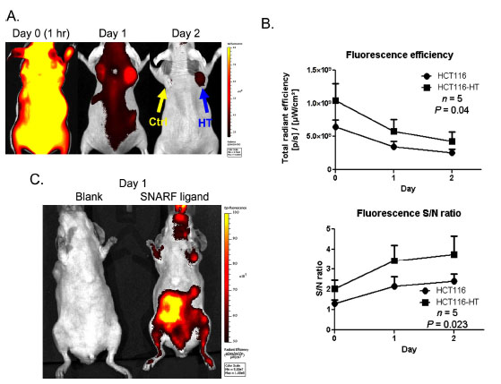
Fig. (4). In vivo uptake of SNARF ligand in HaloTag-expressing tumor xenografts (A) NCr nude mice with HCT116 tumor xenografts (control: left, yellow arrow; HT: right, blue arrow) were intravenously injected with SNARF ligand. Animals were imaged 1h (day 0), 1 day and 2 days after ligand injection with an Ex 640/ Em 700 filter pair. (B) Quantitative presentation and statistical analysis of the imaging data. The upper panel illustrates the fluorescence efficiency of tumors. The lower panel indicates the relative S/N ratios of tumors, normalized using fluorescence of the neck as reference. (C) Non-selective uptake was observed in the gastrointestinal tracks 24h after injection. Control animals received no SNARF ligand injection.