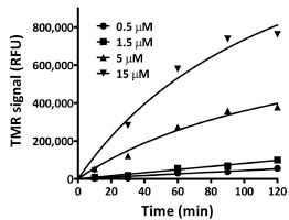
Fig. (3) Time and dose-dependent formation of a stable attachment
between H272F and the TMR-ligand. Plot of fluorescence
intensities (RFU) determined by SDS-PAGE and fluorescence
scanning for TMR. The actual gel image can be found in the
Supplementary Material (Fig. S1A).