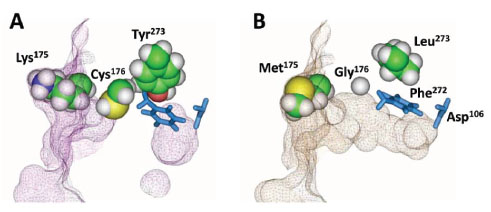
Fig. (6) Structure models of H272F (panel A) and HT2 (panel B) in the absence of bound ligand. The ligand cavities for both variants
were visualized as a Connolly surface using a probe radius of 1.4 Å. HT2 shows a continuous tunnel from the protein surface to the nucleophile,
as opposed to a constricted path indicated for H272F.