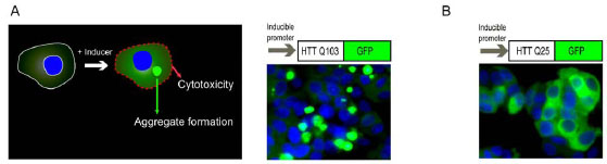
Fig. (1) Schematic representation of a cell based Huntington’s disease model. (A) In a Q103-GFP PC12 cell line, induction of Q103-GFP
fusion protein expression causes formation of protein aggregates and resultant cytotoxicity. Typical screening assays measure either the cytotoxicity
caused by the expression of Q103-GFP fusion proteins or protein aggregates. (B) In a Q25-GFP control cell line, the Q25-GFP fusion
proteins are diffusely distributed in cytosol that do not form aggregates and are not toxic to cells. The images of Q103-GFP and Q25-
GFP in (A) and (B) were obtained with a fluorescence confocal microscope using a 40! objective. Nuclei (blue color) were stained with
Hoechst 33342 and the GFP fusion proteins (either soluble proteins or aggregates) are shown in green color.