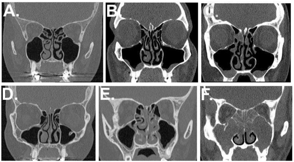
Fig. (1) Evaluation of radiologic signs of nasal polyposis from sinus Computed Tomography (CT) scans. Sinus CT scans that were routinely performed due to clinical purposes. The radiologic NP score was formed based on collected data of the CT scans to evaluation form that was filled by an experienced Radiologist (AM) blinded to patient´s history (12). Sinus mucosal findings were scored as 0=no change, 1=mucosal thickening, 2= polypous mucosal thickening ±discharge. Detectability of turbinate structures was scored as 0=detected; 1= not detectable. The reasons for “not detectable” responses were poor visualization of middle turbinate due to polypoid change or operative modification of turbinate, both considered to be signs of NP. The radiologic NP score 8-16 was suggestive for clinical CRSwNP phenotype. (A) No CT signs of polypous sinus mucosa. Turbinate anatomy is detectable. Radiologic NP score =0. (B) Polypous maxillary sinus mucosa on both sides. No mucosal swelling of anterior ethmoidal cells. Turbinate anatomy is detectable. Radiologic score =4. (C) Polypous maxillary sinus mucosa. Mucosal swelling of anterior ethmoidal cells. Turbinate anatomy is detectable. Radiologic score =6. (D) No polypous sinus mucosa. Right inferior turbinate fine structure is undetectable. Radiologic score =1. (E) Mucosal swelling of right maxillary sinus as well as anterior ethmoidal cells (not shown) and posterior ethmoidal cells. Polypous mucosa of left maxillary sinus. The anatomical fine structure of Inferior and middle turbinates is undetectable. Radiologic score 11. (F) Polypous mucosa of maxillary sinuses and anterior ethmoidal cells (not shown) and posterior ethmoidal cells. The anatomical fine structure of middle turbinates is undetectable. The fine structure of inferior turbinates is detectable. Radiologic score 14.