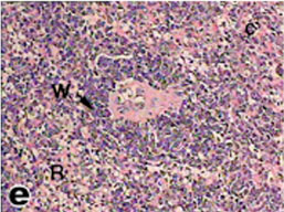
Fig. (13)
Light micrograph of spleen tissue from tortoise showing one white pulp (W) and the rest of the parenchyma is red pulp (R), consisting of cellular cords (c) and numerous venous sinuses. (H & E 400).