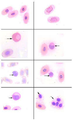
Fig. (2) Light micrograph of Wright's stained blood smears from the desert tortoise Testudo graeca showing: (a) Erythrocytes, (b) Erythrocytes with prominent vacuoles, (c) Heterophil (arrow) and erythrocyte, (d) Eosinophil (arrow) and erythrocyte, (e) Basophil (arrow) and erythrocyte, (f) Lymphocyte (arrow) and erythrocyte, (g) Monocyte (arrow) and erythrocyte, (h) Clumps of thrombocytes (arrows) and erythrocyte. (Wright stain 1000).