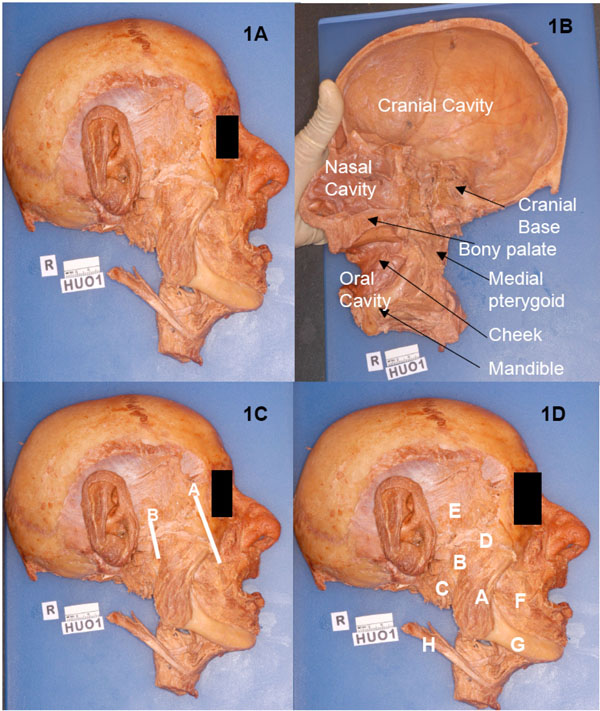
Fig. (1) A) Specimen HU O1, the outer aspect of the right half head. B) Specimen HU O1, the medial aspect of the right half head. C)Specimen HU O1, lines of osteotomy to mobilize the zygomatic arch. D) Specimen HU O1, significant bones and muscles on the lateral aspect of the specimen, showing the superficial part of the masseter (A), the deep part of the masseter (B), the parotid gland (C), the zygomatic arch (D), the temporalis muscle and fascia (E), the buccinator muscle (F), the mandibular body (G) and the posterior belly of the digastric (H).