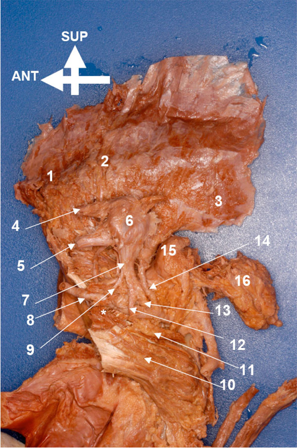
Fig. (4) Specimen HU O1, medial aspect of the masticatory
muscles and the mandibular nerve complex, right block (*indicates
the inferior belly of the lateral pterygoid muscle). 1, Temporalis
(pars superficialis). 2, Temporalis (pars profunda, main portion). 3,
Temporalis (pars profunda posterior). 4, Ophthalmic nerve (CN
V1). 5, Maxillary nerve (CN V2). 6, Trigeminal ganglion. 7,
Mandibular nerve (CN V3; main stump). 8, Maxillary artery
(pterygoid section). 9, Buccal nerve. 10, Medial pterygoid muscle
(main body). 11, Medial pterygoid muscle (upper part). 12, Lingual
nerve. 13, Stump of inferior alveolar and auriculotemporal nerves.
14, Middle meningeal artery. 15, Head of condyle (covered by
disc). 16, Parotid gland.