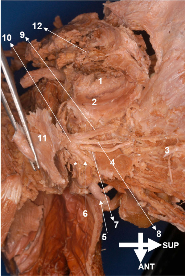
Fig. (5) Specimen HU O1, superior aspect of the masticatory
muscles and the mandibular nerve complex, left block (*indicates
the inferior belly of the lateral pterygoid muscle). 1, Head of
condyle covered by disc. 2, Disc-superior lateral pterygoid junction.
3, Temporalis (deeper aspect). 4, Superior lateral pterygoid (upper
view). 5, Maxillary artery (pterygoid section). 6, Buccal nerve. 7,
Middle deep temporal nerve. 8, Posterior deep temporal nerve. 9,
Nerve to the masseter (main stump). 10, Superior division of
mandibular nerve. 11, Trigeminal ganglion. 12, Parotid gland.