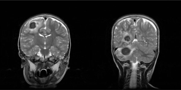
Fig. (2)
T -2 weighted magnetic resonance imaging of the brain of case #1, showing some of the intraparenchymal TB abscesses.