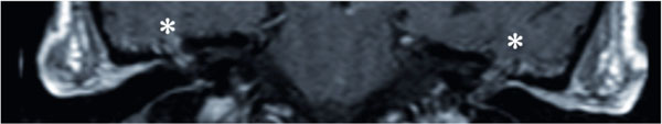
Fig. (7)
T1-weighted coronal MRI with gadolinium of the temporal bones and skull base of case #6. Aterisk (*) indicates the areas of bilateral encephaloceles herniating through the middle cranial fossa floor.