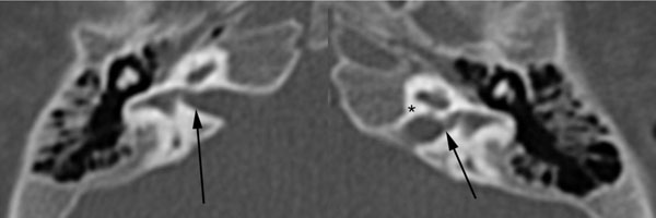
Fig. (8)
Axial computed tomography of the temporal bones of case #7 showing bilateral widening of the canals for the superior vestibular nerves (solid black arrows). Asterisk (*) is directly adjacent to the left auditory nerve canal.