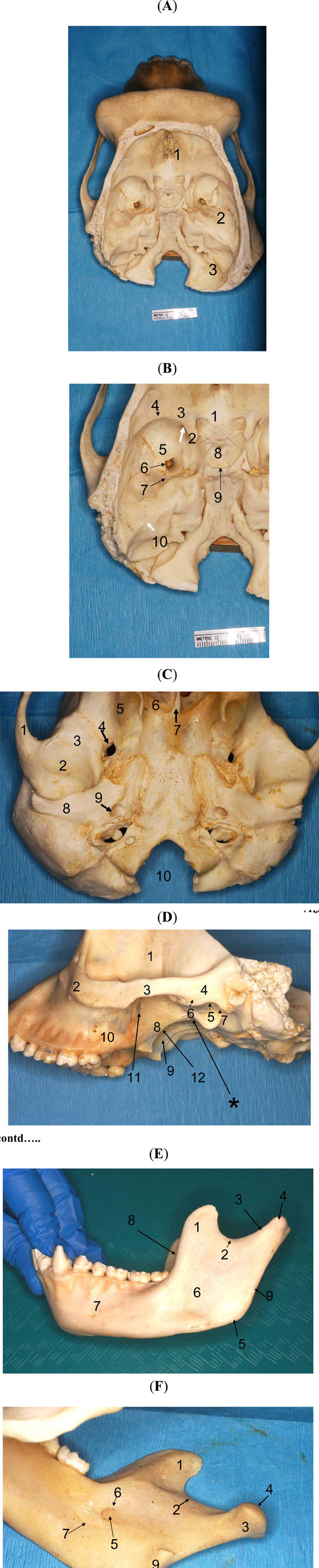
Fig. (2) Pan troglodytes, dry skull. A, Basicranium, internal aspect,
general view (1, anterior cranial fossa; 2, middle cranial fossa; 3,
posterior cranial fossa). B, Basicranium, internal aspect, detailed
view of the middle and posterior cranial fossae (1, chiasmatic
sulcus; 2, anterior clinoid process ; 3, lesser wing of sphenoid ; 4,
spheno-parietal eminence ; 5, greater wing of sphenoid ; 6, foramen
ovale ; 7, petro-sphenoid suture ; 8, sella turcica ; 9, dorsum sellae ;
10, superior petrosal eminence; the white arrows points towards the
superior orbital fissure and the foramen rotundum). C, Basicranium,
external (inferior) aspect (1, zygomatic process of temporal; 2,
mandibular fossa; 3, articular tubercle; 4, foramen ovale; 5,
pterygoid fossa; 6, posterior nasal opening; 7, vomer; 8, postglenoid
eminence; 9, carotid canal; 10, foramen magnum). D,
basicranium, lateral aspect, left side (1, temporal fossa, 2,
zygomatic bone; 3, zygomatic arch; 4, temporal-zygomatic process;
5, mandibular fossa; 6, articular eminence; 7, post-glenoid
eminence; 8, lateral pterygoid plate; 9, medial pterygoid plate; 10,
posterior wall of maxilla; 11, pterygo-maxillary fissure; 12,
pterygoid fossa; the asterisc points towards the foramen ovale). E,
mandible, lateral aspect, left side (1, coronoid process; 2,
mandibular notch; 3, neck of condyle; 4, head of condyle; 5, angle
of mandibular ramus; 6, masseteric fossa (mandibular ramus); 7,
mandibular body; 8, mandibular ramus-anterior margin; 9,
mandibular ramus-posterior margin). F, mandible, medial aspect,right side (1, coronoid process; 2, mandibular notch; 3, neck of
condyle; 4, head of condyle; 5, mandibular foramen; 6, lingular
process; 7, mylohyoid groove; 8, medial pterygoid tubercle; 9,
stylomandibular tubercle).