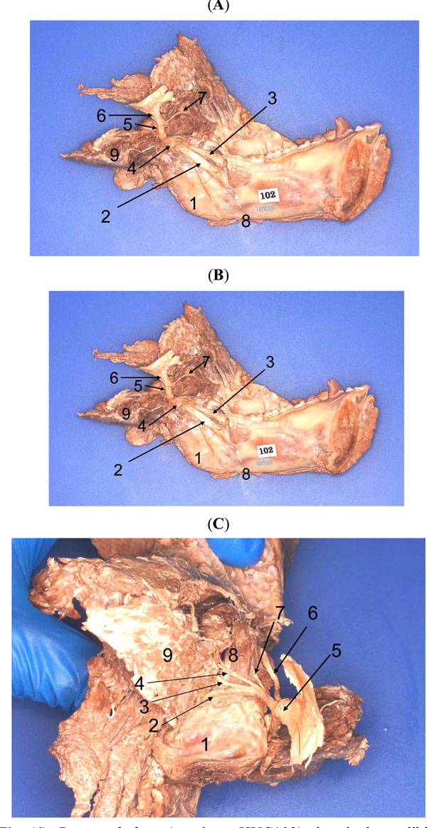
Fig. (6) Pan troglodytes(specimen HUC102), detached mandible.
A, Medial aspect, left side (1, trigeminal nerve-main stump; 2,
trigeminal ganglion; 3, ophthalmic nerve; 4, maxillary nerve; 5,
mandibular nerve; 6, buccal nerve; 7, superior head of lateral
pterygoid; 8, inferior head of lateral pterygoid; 9, tensor veli
palatini; 10, medial pterygoid; 11, nerve to medial pterygoid; 12,
nerve to tensor veli palatini; 13, auriculo-temporal nerve; 14,
maxillary artery; 15, external carotid artery; 16, superficial
temporal artery; 17, parotid gland (medial aspect); 18, temporalis 19, head of condyle). B, Medial aspect, left side, showing major
branches of V3 (1, mylohyoid nerve; 2, inferior alveolar nerve; 3,
lingual nerve; 4, chorda tympani nerve; 5, mandibular nerveposterior
division; 6, mandibular nerve-main stump; 7, buccal
nerve; 8, mandibular foramen; 9, medial pterygoid [lifted up]). C,
Superior aspect showing the branches of the anterior division of V3
(1, TMJ disk over the condyle; 2, nerve to masseter; 3, deep
temporal nerve; 4, deep temporal nerve; 5, mandibular nerveanterior
division; 6, buccal nerve; 7, nerve to superior lateral
pterygoid; 8, superior lateral pterygoid; 9, temporalis).