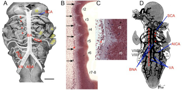
Fig. (14) Rhombomeric transverse branches of basilar. A. The vertebro-basilar arterial pattern of non-human primates is similar to that of
humans. This rhesus monkey (Macaca mulatta) hindbrain modified after Gerhard and Olszewski [100] shows a midline unpaired basilar
artery (BA) giving off multiple symmetrical transverse branches. The segmental correlation of brain vasculature and neuronal development
can be seen as the abducens nerve (VI), located in r5, can also be demonstrated by counting down 4 transverse branches (red asterisks) from
the superior cerebellar artery (SCA) to rhombomere 5. B. Sagittal section of Carnegie Stage 14 Human Embryo (# 9297) showing
rhombomere borders and sub-pial vascular elements forming the pontine branches of the basilar artery (arrows). C. Higher magnification of
early transverse vessel (*) lying adjacent to pial surface of r5-6 inter-rhombomeric border. D. Ventral view of graphic reconstruction
(modified from Padget [14]) of BA forming by fusion of the primitive bilateral neural arteries (BNA). Transverse arteries (red asterisks)
clearly arise prior to this fusion. Red line shows plane of section in B. III, oculomotor nerve; V, trigeminal nerve; VI, abducens nerve; VII,
facial nerve; VIII, vestibulocochlear nerve; AICA, anterior inferior cerebellar artery; ASA, anterior spinal artery; PICA, posterior inferior
cerebellar artery; VA, vertebral artery.