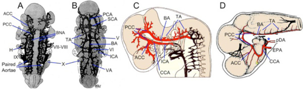
Fig. (3) Development of Carotid-Basilar Axis in Human and Dogfish A-B. Graphic reconstructions of ventral views of the brain and
cranial arteries in Stage 13 (4mm; A) and Stage 16 (9mm; B) human embryos, modified from Padget [14]. By 4mm, the internal carotid
artery (ICA) has already divided into cranial (anterior; ACCs) and caudal (posterior; PCCs) divisions. Note that the bilateral neural arteries
(BNAs) and adjacent pial plexus reorganize between these two stages to form a vertebro-basilar system that has the same basic topography as
in adults. C. Lateral view of reconstructed 9mm (stage 16) human embryo modified from Padget [14] showing the cerebral carotid artery
(CCA) passing caudal to the optic vesicle and dividing into ACCs and PCCs. The basilar artery (BA) has been formed from the fusion of the
BNA and already is giving off transverse arterial branches (TA). D. Lateral view of the brain and carotid branches of a 32 mm embryo of
Squalus acanthias modified from Sterzi [45]. The paired dorsal aorta (pDA) join to form an unpaired “encephalic” trunk (asterisk) which
then enters the hypophyseal fossa and divides into left and right cerebral carotids (CCAs) which are joined by the efferent pseudobranchial
arteries (EPAs). The CCA divides into ACC and PCC divisions, with the latter curving caudally beneath mid and hindbrain where they fuse
in the midline to join the BA. Cranial nerve roots are indicated by roman numerals as H, hyoid artery; PCA, posterior cerebral artery; SCA,
superior cerebellar artery; VA, vertebral artery.