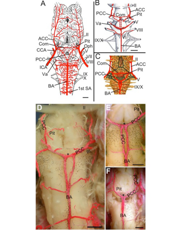
Fig. (9) Amphibians (Salamander and Frog). A. Cerebral vasculature of Ambystoma tigrinum modified after Roofe [76] showing the
cerebral carotid arteries (CCAs) giving off ophthalmic arteries (Ophs) and dividing into anterior (ACC) and posterior (PCC) divisions. The
PCCs give off pituitary branches (Pit), form a short crossbridge (Com), and then join (red dotted line) to form a midline unpaired basilar
artery (BA) that gives off numerous transverse branches, including large vessels traveling with trigeminal (Va) and vestibulocochlear nerves.
B. Illustration of Salamandra salamandra modified after Francis [24] showing the unification of the PCCs to form a crossbridge (Com; red
dotted lines) and giving off two parallel caudal branches (red dotted lines) that join to form an unpaired BA. Pituitary (Pit), trigeminal (Va)
and vestibulocochlear branches are also shown. C. Illustration of Salamandra maculosa modified after Schöbl [23] showing a third variation
of the unpaired BA formation that combines features seen in A and B. No magnification was given by Schöbl for C. D-F. In Rana
catesbiana, although the PCC travel caudally and fuse to form the BA, the length they travel paired varies in different specimens. The PCC
give off pituitary and mesencephalic branches, form a short crossbridge (*), and then travel caudally before fusing to form an unpaired BA.
All specimens had symmetrical transverse branches coming off the BA. A partially fused BA with a paired region (fenestration) just caudal
to the PCC crossbridge (F) was also found. II, optic nerve; V, trigeminal nerve; VII, facial nerve; VIII, vestibulocochlear nerve; IX,
glossopharyngeal nerve; X, vagus nerve. Scale bars are 1mm in A-B and 1mm in D-F.