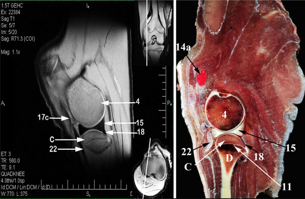
Fig. (1) Sagittal MR image (A) and gross anatomic section (B) of the left buffalo stifle at the level of 3 cm lateral to the Fossa
intercondylaris (dorsal is up and caudal is to the right of the viewer). A, Corpus ossis femoris; B, Distal epiphysis of os femoris;
C, Extremitas proximalis tibiae; D, Corpus tibiae; 1, Trochlea ossis femoris (1a, medial ridge; 1c, intertrochlear groove); 2, Facies popliteal;
3, Condylus medialis of os femoris; 4, Condylus lateralis of os femrois (4a, Fossa extensoria); 6, Patella (6a, Basis patellae; 6b, Processus
cartilaginous; 6c, Apex patellae; 6d, Facies articularis; 6e, Facies cranialis); 7, Condylus lateralis of the tibia (7a, Incisura Poplitea);
8, Tuberositas tibiae; 9, Distal epiphysis of the femur ossification center; 10, Tibial tuberosity ossification center; 11, Proximal diaphysis of
the tibia ossification center; 12, medial femorotibial sac; 13, lateral femorotibial sac; 14, femoropatellar synovial sac (14a, proximal pouch);
15, Meniscus lateralis (15a, cranial meniscotibial ligament; 15b, Lig. Meniscofemorale); 16, Meniscus medialis (16a, cranial meniscotibial
ligament); 17, Ligg. Cruciata genus (17a, Lig. cruciatum craniale; 17b, Lig. cruciatum caudale); 18, Lig. popliteum obliquum;
19, Fibrocartilagines parapatellaris medialis; 20a, Lig. patellae intermedium; 20b, Lig. patellae mediale; 20c, Lig. patellae laterale; 21,
Corpus adiposum infrapatellare; 22, Common tendon of the extensor digitorum longus and peronaeus tertius; VM, M. vastus medialis;
VI, M. vastus intermedius; GL, M. gastrocnemius (Caput laterale);GM, M. gastrocnemius (Caput mediale); SD, M. flexor digitorum
superficialis; POP, M. popliteus; ST, M. semitendinosus; SM, M. semimembranosus.