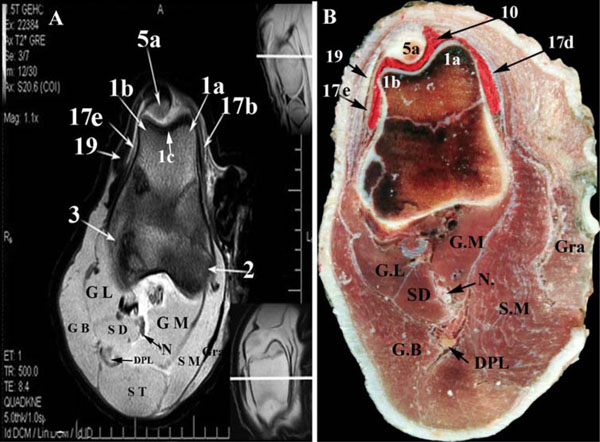
Fig. (4) Transverse MR image (A) and gross anatomic section (B) of the left buffalo stifle at the level of the patellar apex (dorsal is up and
lateral is to the left of the viewer). 1, Trochlea ossis femoris (1a, medial ridge; 1b, lateral ridge; 1c, intertrochlear groove); 2, Condylus
medialis of os femoris; 3, Condylus lateralis of os femoris; 3a, Fossa extensoria; 4, Fossa intercondylaris; 5a, Apex patellae; 6, Eminentia
intercondylaris (6a, tuberculum intercondylare mediale; 6b, tuberculum intercondylare laterale); 9, medial femorotibial synovial sac;
10, femoropatellar synovial sac; 11, Meniscus Lateralis (11a, cranial meniscotibial ligament of the lateral meniscus; 11b, Lig.
Meniscofemorale of the lateral meniscus); (12, Meniscus medialis (12a, cranial meniscotibial ligament; 12b, caudal meniscotibial ligament);
13a, Lig. cruciatum craniale; 13b, Lig. cruciatum caudale; 14, Lig. collaterale laterlae; 15, Lig. collaterale mediale; 16, Lig. popliteum
obliquum; 17a, Lig. patellae intermedium; 17b, Lig. patellae mediale; 17c, Lig. patellae laterale; 17d, Lig. femoropatellare mediale; 17e,
Lig. femoropatellare laterale; 18, Corpus adiposum infrapatellare; 19, Gluteobiceps tendon; 20, Common tendon of the extensor digitorum
longus and peronaeus tertius; T, proximal Extremity of the tibia; DPL, Deep popliteal lymph node; GL, M. gastrocnemius (Caput laterale);
GM, M. gastrocnemius (Caput mediale); SD, M. flexor digitorum superficialis; POP, M. popliteus; GB, M. gluteobiceps femoris; ST,
M. semitendinosus; SM, M. semimembranosus; Gra, M. gracilis.