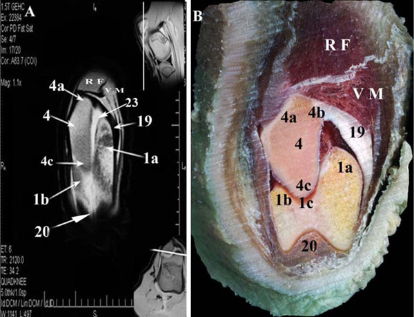
Fig. (8) Dorsal MR image (A) and gross anatomic section (B) of the left buffalo stifle 2cm caudal to the most cranial part of trochleae ossis
femoris (dorsal is up and lateral is to the left of the viewer). 1, Trochlea ossis femoris (1a, medial ridge; 1b, lateral ridge; 1c, intertrochlear
groove); 2, Condylus medialis of os femoris; 3, Condylus lateralis of os femoris (3a, Fossa extensoria); 4, patella; (4a, Basis patellae; 4b,
Processus cartilagineus; 4c, Apex patellae); 5, Condylus medialis of the tibia; 6, Condylus lateralis of the tibia; 7, Eminentia intercondylaris
(7a, tuberculum intercondylare mediale; 7b, tuberculum intercondylare laterale); 8, Tuberositas tibiae; 10, Distal epiphysis of the femur
ossification center; 11, Proximal diaphysis of the tibia ossification center; 12, lateral femorotibial sac (12a, distal pouch); 13, Meniscus
lateralis; 14, Lig. Meniscofemorale; 15, Meniscus medialis (15a, caudal meniscotibial ligament); 16a, Lig. cruciatum craniale; 16b, Lig.
cruciatum caudale; 17, Lig. collaterale laterlae;18, Lig. popliteum obliquum; 19, Fibrocartilagines parapatellaris medialis; 20, Lig. patellae
intermedium; 21, Common tendon of the extensor digitorum longus and peronaeus tertius; 22, Popliteal artery and vein; 23, Femoropatellar
joint; VM, M. vastus medialis; RF, M. rectus femoris; GL, M. gastrocnemius (Caput laterale); GM, M. gastrocnemius (Caput mediale); SDF,
M. flexor digitorum superficialis; GB, M. gluteobiceps femoris.