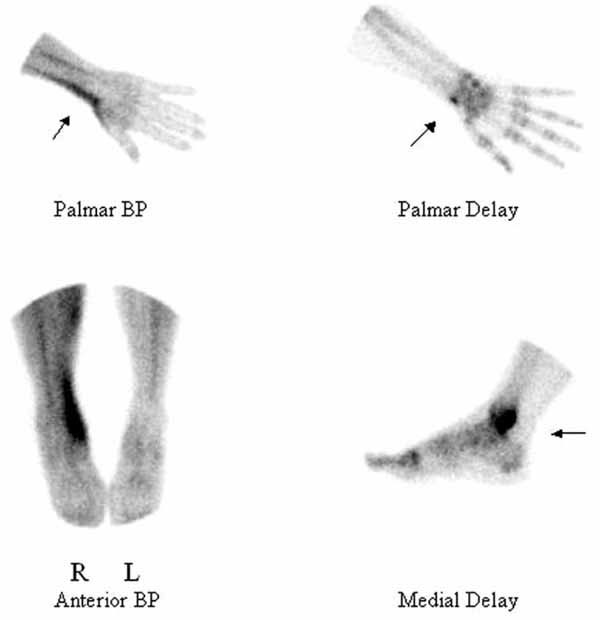
Fig. (1) Upper panel shows bloodpool (BP) and delayed static (Delay) views of right hand. Note linear hyperemia along the radial wrist at
the tendon sheath, and a slightly increased focal skeletal uptake in the right radial styloid, demonstrating tenosynovitis. Lower panel shows
bloodpool (BP) and delayed static (Delay) views of right foot. Note the curvilinear band of increased blood pool activity in the right medial
ankle, corresponding to the anatomic position of the tibialis posterior tendon (left panel), and an intensely increased focal skeletal uptake in
the right dorsomedial ankle, not peri-articular (right panel), demonstrating tibialis posterior tendinitis without arthritis.