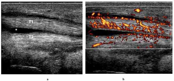
Fig. (2) a. Longitudinal grey-scale ultrasonographic scan of the medial aspect of the right ankle showing hypoechoic fluid surrounding the hyperechoic tendon of the tibialis posterior tendon. b. shows a power Doppler sonography scan of the tendon of the tibialis posterior muscle.