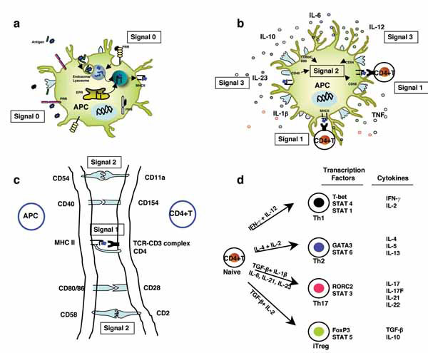
Fig. (1) (a) Immature DCs present with high capacity to take up exogenous antigens. The recognition of antigen-associated PAMPs by PRRs (signal 0) leads to maturation and activation of DCs. In endosomes/lysosmes, the internalized antigens are processed into smaller peptides. These antigenic peptides bind to MHC II molecules and transported to cell surface. EPR, endoplasmic reticulum (b) The ‘antigen-educated’ DCs present antigenic peptides in the context of MHC II to TCR of naïve CD4+T cells (Signal 1). These mature and activated DCs express high levels of MHC, co-stimulatory (CD40, CD80 and CD86) and adhesion molecules (CD54 and CD58) (signal 2) and secrete high amounts of cytokines IL-1α, IL-6, IL-10, IL-12, IL-23 and TNF (Signal 3) that play a critical role in inflammation and differentiation of CD4+T cells. (c) The interaction of signaling molecules at APC-T cell interface (d) CD4+T cells under the influence of signaling by DCs and DC-derived cytokines can be differentiated into 4 distinct subclasses expressing distinct transcription factors: Th1, Th2, Th17 and induced Tregs (iTregs). The figure is reprinted from Trends Pharmacol Sci 2009, 30:287-295 (ref. [10]) with permission from Elsevier.