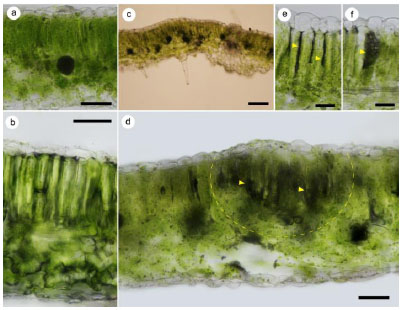
Fig. (2)
Transverse sections of healthy (a, b) and C. michiganensis infected (c-f) leaves: a - healthy leaf; b – healthy leaf after injection of sterile water; c, d - leaf of the infected plant showing initial signs of bacteriosis (the zone of the columnar mesophyll is outlined, where the induced protective reaction develops; arrows show necrosis) e - transformation of the cell walls of the columnar mesophyll in infected leaf; f - necrotized mesophyll cell in infected leaf; bar: a - 50 μm; c - 80 μm; b- 40 μm, e, f - 20 μm.