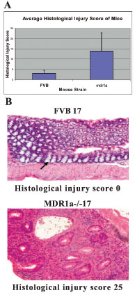
Fig. (2) A)- Graphical representation of the average histology
scores of the mice. This graph shows the averages and error bars
for the two mouse strains FVB control mice and mdr1a -/- gene
targeted mutant mice. B)- Cross sectional images of colonic tissue
from an FVB control mouse (histological injury score of 0) and an
mdr1a -/- gene targeted mutant mouse (inflammatory bowel
disease model with histological injury score of 25). The FVB
mouse colonic tissue contains crypts (example indicated by black
arrow), which are columns of white goblet cells and are normal
structures of a healthy gut, with no cells between them. However
the mdr1a -/- gene targeted mutant mouse colonic tissue contains
very few crypts with no visible goblet cells inside. Almost all of
the structures of the mucus layer are destroyed and the crypt
cavities begin to fill with pink pus cells. The purple dots located
between the cavities are inflammatory cells.