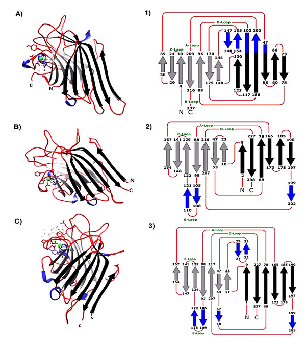
Fig. (1)
A, B and C are the monomers of Con A, ECorL and ECL crystallized with hapten ligand [pdb files: 1c57, 1axy and 1gzc respectively]. The 3D- monomer structures were opened and edited using UCSF-Chimera 1.8 software [1-3]. are the topology diagram of each lectin deduced from the 3D-structure from each lectin. Black arrows represent the β-back black sheets, the light gray arrows represent the β-front sheets and the small blue arrows are the S-sheets while the red lines represent the loops. A, B, C, and D-loops contains the amino acids responsible for sugar binding.