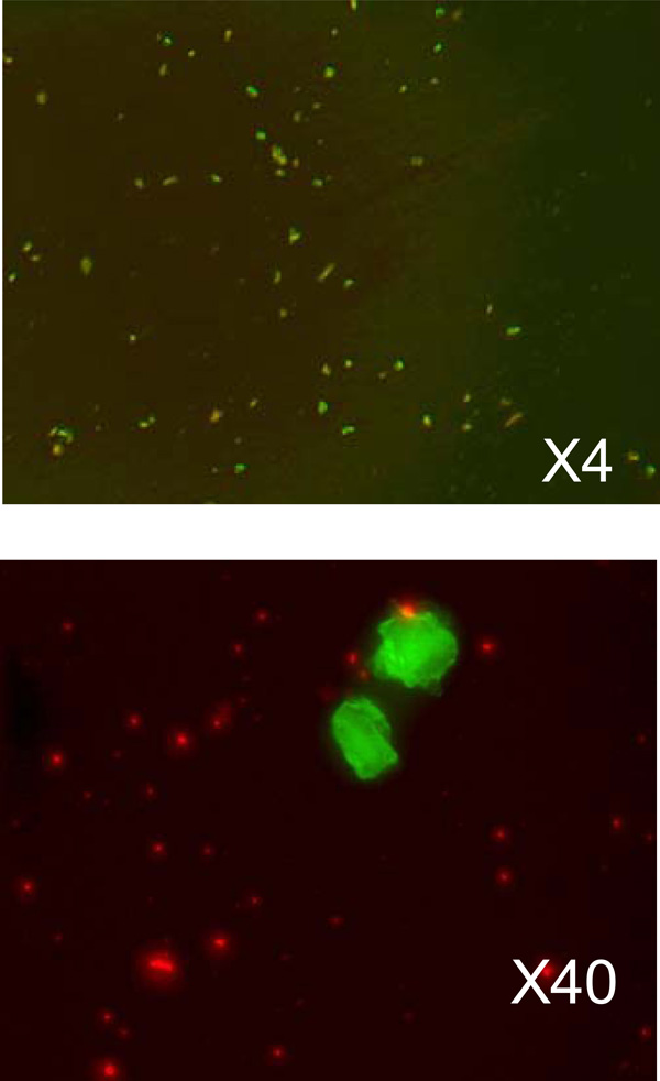
Fig. (4) The microscopic images of transfected cancer cells. The upper image is taken with 4-times magnification and show the overall density of the transfected cells. The lower image shows transfected cells in more detail. Green fluorescence is from GFP, and red fluorescence - from liposomes.