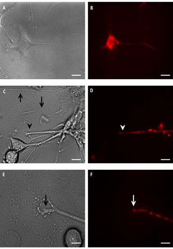
Fig. (1)
Time lapse videomicroscopy. Panels A and B show a PC12 cell body and a neurite under bright field (A) and fluorescence conditions (B). At low magnification under bright field (C) and fluorescence conditions (D), neurites can be seen taking the direction of the groove (arrows). Arrow heads on panel C and D point a neurite where fluorescent mitochondria are observed. On Panel E (Bright field) and F (Fluorescence), higher magnification shows the growth cone of a neurite and fluorescent mitochondria (arrow). Scale bar is 10µm on A, B, C and D. and 5µm on E and F.