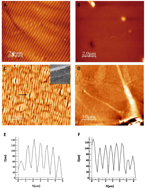
Fig. (3)
AFM analysis of the surface azopolymer pattern before (panel A) and after applying a usual preparation of surfaces for cell growing (panel B). On panel B, the network has been erased. Using our protocol, the surface pattern was preserved (panel C) with the same width (around 1µm, inset in panel C). A neuronal cell can be visualized on the azopolymer surface pattern (panel D) showing the possibility to grow cells with our protocol.